Confocal microscopy at its full potential
Achieve imaging precision with our advanced light loss correction technology for confocal microscopy
The Unmet Need in 3D Sample Imaging
Confocal fluorescence microscopy is the gold standard for imaging 3D samples like organoids, spheroids, and tissues.
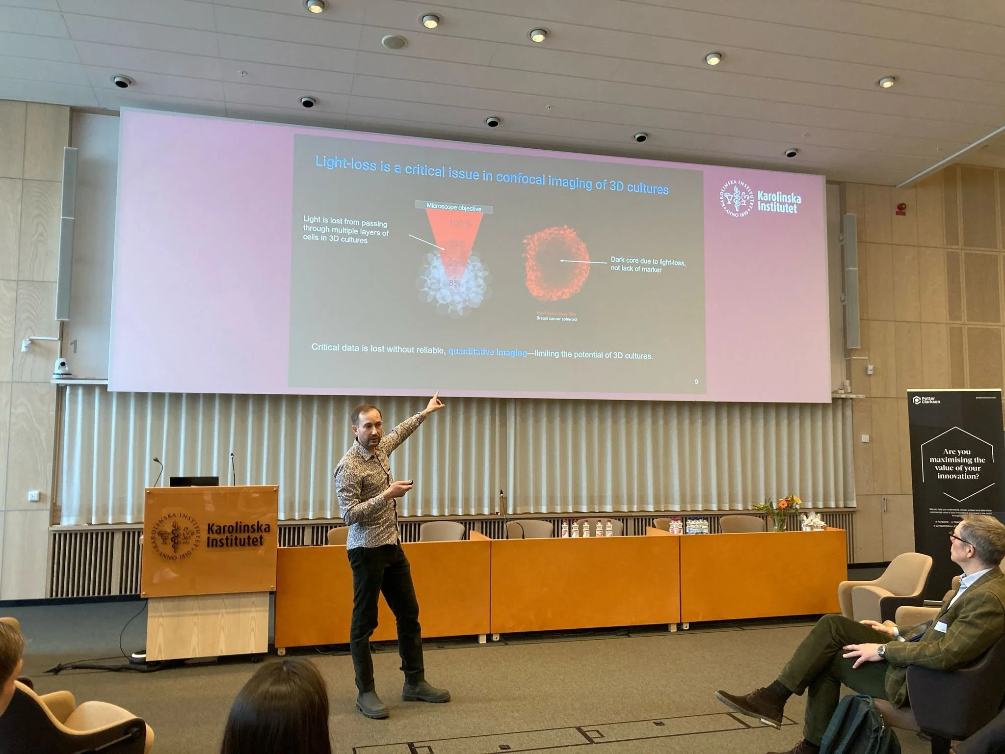
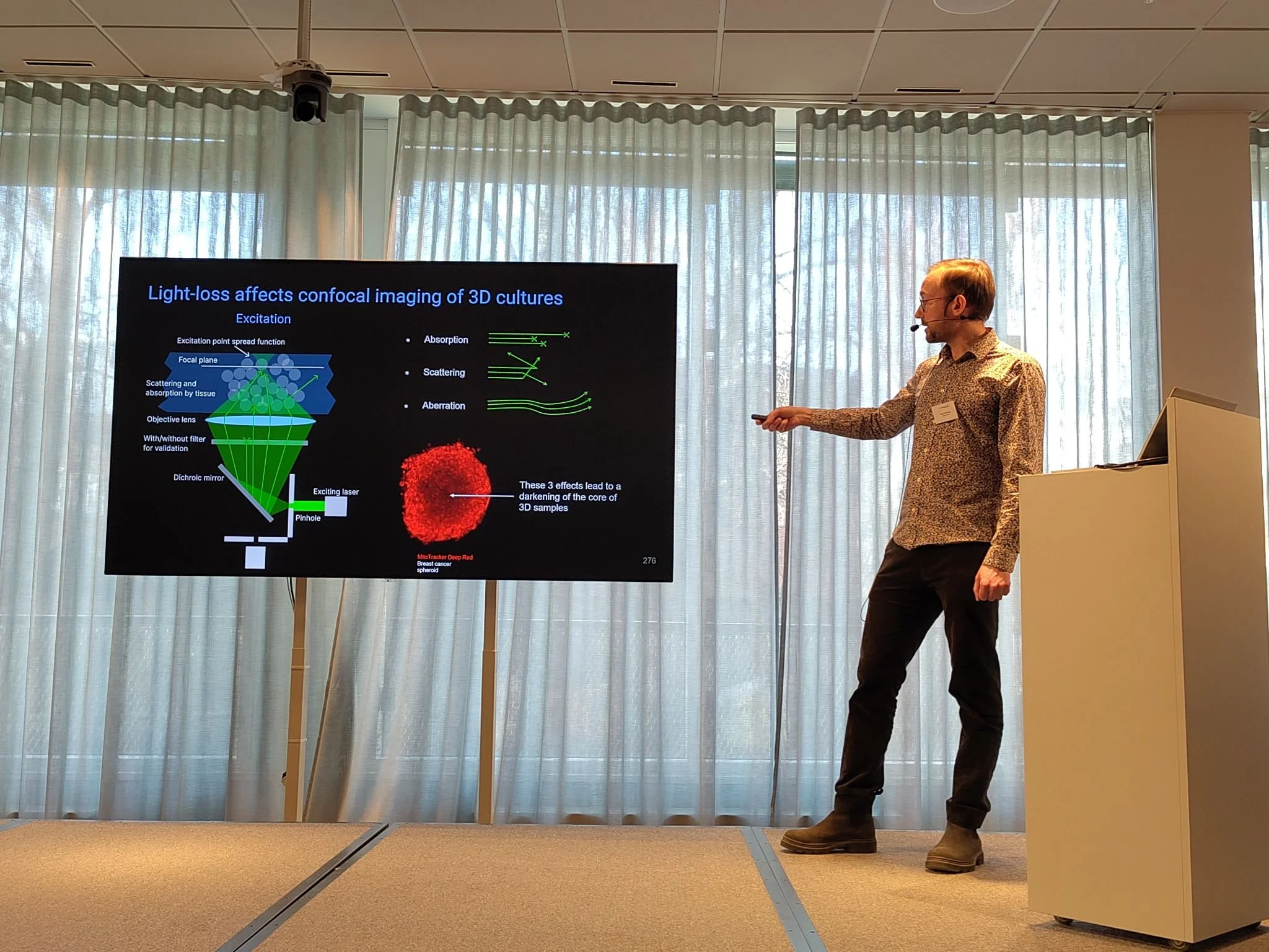
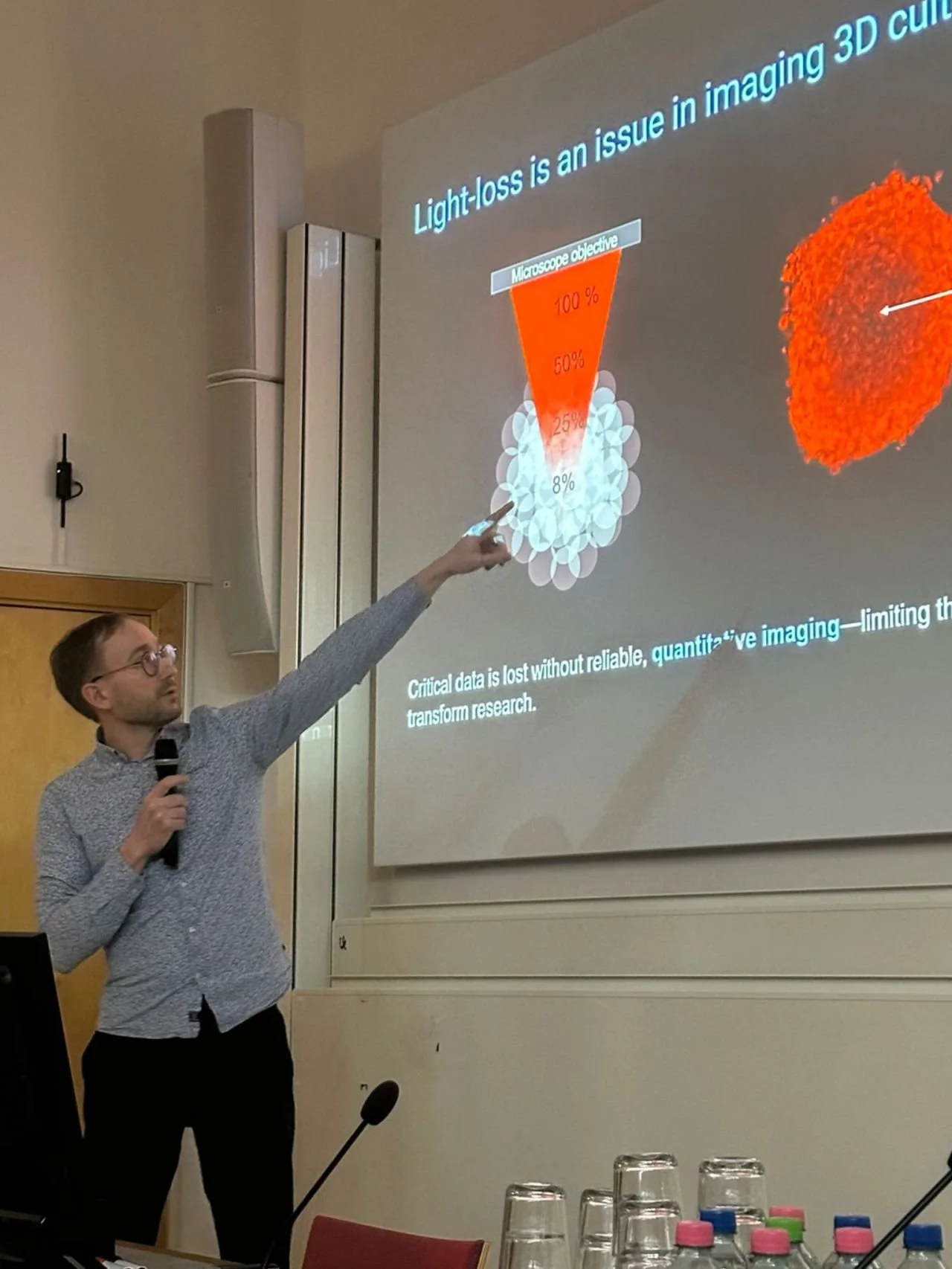
However, these 3D samples when thick enough, cause light absorption and scattering, leading to distorted images and unreliable results.
Yet today, no solution fully addresses this issue, preventing researchers from accurately characterizing deeper structures in 3D samples using confocal microscopy.
Uncertain Signal Interpretation
Without a clear way to differentiate real biological absence from technical signal distortion, scientific conclusions remain uncertain.
Inconsistent Quantification
Signal loss at deeper layers skews fluorescence intensity, making it hard to compare results across depths or samples.
Our Solution
Jellyfish
Our solution is a patent-pending imaging correction technology, designed to improve the accuracy of confocal microscopy for 3D samples like organoids, spheroids, and tissues.

Jellyfish Software uses a focus model fit to pixel-level measurements to correct signal attenuation in z-stacks.
The current prototype runs as a standalone software, with the option for automated use through a Python API.

-
See the true fluorescence signal without distortion
-
From confocal images to spatial fluorophore data
-
Distinguishing real biological effects from artifacts
The Science Behind Ciprocity
Challenge
Light Absorption & Scattering
Thick 3D samples distort imaging, making it unclear whether signal loss is due to biological factors or imaging limitations.
Innovation
Physics-Based Correction
By measuring the amount of absorption and scattering of light, we correct for light loss effects, ensuring that fluorescence signals reflect actual biological markers.
Breakthrough
Quantitative Confocal Imaging
For the first time using confocal microscopy, our technology enables measurement of spatial differences in fluorophore concentration.
RECENT NEWS
-
TALKS
AND
CONFERENCES
-
RECENT NEWS - TALKS AND CONFERENCES -
Recent News
-
Awarded SEB Innovation Scholarship 2025
April 2025
We were 1 among the 10 selected teams in Sweden for the SEB Innovation Scholarship 2025 during KI Innovations' Investor Day.
-
New Collaborators
We’ve been—and continue to be—engaged in research collaborations with imaging experts and core facilities worldwide, including in Sweden, Norway, Finland, Spain, and New Zealand. These partnerships are key to validating our technology across diverse biological samples and imaging environments. -
Part of the DRIVE Life Science Incubator
Since 2024, we’ve been part of the DRIVE program at KI Innovations, and is being instrumental in accelerating our journey from concept to product.
Talks and Conferences
-
![]()
KI Innovations InvestorDay 2025
April 2025
At KI Innovations' Investor Day, we pitched our solution to a room full of investors and connected with visitors at our booth.
-
![]()
Lab & Diagnostics of The Future
March 2025
We presented our technology at the Lab & Diagnostics of the Future conference, sharing how we measure and correct light-loss in confocal imaging.
-
![]()
CATALYZER program Final
January 2025
We wrapped up the Catalyzer program at KI Innovations by pitching Ciprocity at the Dragons’ Den finale. It was a great opportunity to share our vision, receive feedback from industry experts, and connect with investors.
-
![]()
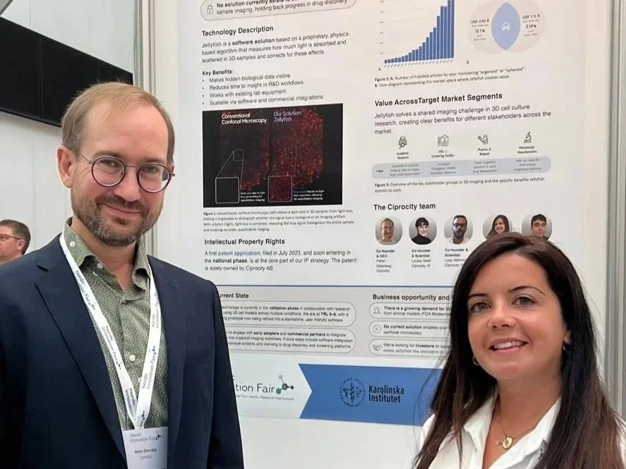
Nordic Innovation Fair 2025
September 2025
Our team members Petter Säterskog and Marina Polo Gozalo represented Ciprocity at the Nordic Innovation Fair 2025, showcasing our technology during the poster session, pitching in 1:1 meetings, and connecting with innovators across the Nordic life science ecosystem.
-
![]()
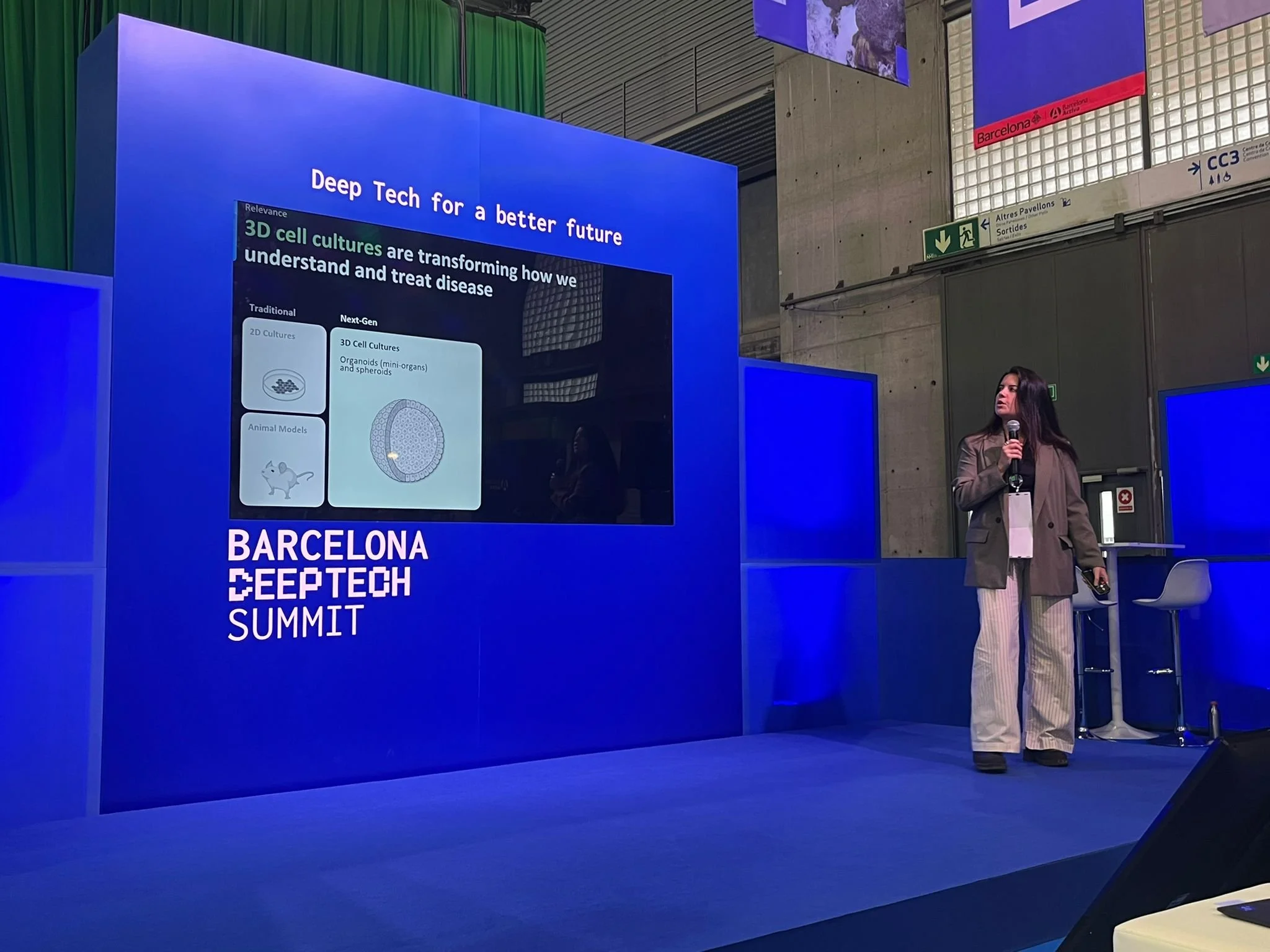
Barcelona Deep Tech Summit 2025
November 2025
We were selected as one of 10 startups to join the Stockholm Business Region delegation at the Barcelona Deep Tech Summit 2025. Our Business Developer Marina Polo Gozalo represented Ciprocity in our booth, pitching our solution and sharing how Jellyfish supports researchers.
FAQs
-
Jellyfish is currently offered as a prototype desktop application run by the Ciprocity team to our collaborators. We're building a beta testing program and plan to offer full user support, onboarding, and documentation as we scale.
-
We are currently validating Jellyfish across different 3D sample types including organoids and spheroids·. Early testing shows consistent signal restoration across samples and imaging conditions.
-
Yes. Jellyfish is designed for researchers with minimal technical background, offering a simple desktop interface for Mac and Windows.
-
No. Jellyfish runs as a standalone desktop app for Mac and Windows, enabling image correction anywhere.
About Us
Our Story
Our adventure started in 2023, when trying to address a recurring problem in our research projects using spheroid imaging: the gold standard for 3D sample imaging wasn't allowing for quantitative marker analysis.
We are now aiming to improve the reliability of microscopy by releasing a tool to analyze confocal images.
The Team
A team of enthusiastic researchers striving for better science, solving problems we've faced ourselves.
CEO
Optical Modeling & Algorithm Design
Dr. Säterskog has obtained his PhD in theoretical physics from Leiden University and has since continued research both in theoretical physics at NORDITA and in cancer biology at Karolinska Institutet which lead to the innovation Ciprocity's product Jellyfish is based on. Apart from academic research, Dr. Säterskog has experience developing software in the biomedical industry.
Organoid Expert
Organoid generation & imaging
Loay is a PhD student in Bioengineering at NTNU. He has a background in Pharmacy and worked as a clinical pharmacist in pediatric oncology for three years. He then moved to Sweden to pursue a master's degree in Experimental and Medical Bioscience, studying at both Linköping University and Karolinska Institute. His work with Ciprocity is primarily experimental, focusing on microscopy.
Marina Polo Gozalo
Antony Cougnoux
BD & Marketing Manager
Business Development & Marketing Communications
Marina has a background in Biochemistry (BSc) and Biomedical Research (MSc), with experience in academic research labs, including as a technician at KI's flow cytometry core facility. Graduated from Bioentrepreneurship MSc at KI, she connects science and business at Ciprocity. Before, she has supported several early-stage life science startups as a Business Consultant and gained corporate experience in both Sweden and New Zealand.
Scientific Advisor
Scientific Support & Protocol Design
Antony has 10+ years of experience working across fields of biology. Since graduating with a PhD in Microbiology he worked in cancer biology and neuroscience. His work involves extensive microscopy use, especially confocal. He is currently working for The Hide & Seek Foundation exploring therapeutic avenues for a rare pediatric disease.
Louise Gsell
Bioinformatician
Development Strategy & Software Testing
Louise is a computational and experimental Ph.D. student in cancer immunology who has experience developing computational tools for a bioanalytical organization and in clinical-stage oncology-focused biotech companies. Her current work involves an important part of communication between biologists and computer scientists, she is part of the image analysis teams of Ciprocity and assists the collaborators in utilizing the prototype.

Loay Mahmoud
Petter Säterskog
Contact Us
We’re open to collaborations, pilot projects, and investment discussions.
Fill in the form or reach out to us at:
For Researchers
For Investors

Our Vision
Transforming confocal microscopy from imaging to
quantitative measurements
Sponsors & Partners